 Español
 English
 Deutsch
 Italiano
 Português
- Auteur:
- Eduardo Anitua
- Gorka Orive
- Miriam Idoipe
- Borja de la Sen-Corcuera
- Ronald M Sánchez-Ávila
- Carmen Sánchez-Pérez
- María Satué
- Antonio Sánchez-Pérez
- Francisco Muruzabal
- Luis Pablo
Membrane of Plasma Rich in Growth Factors in Primary Pterygium Surgery Compared to Amniotic Membrane Transplantation and Conjunctival Autograft
Abstract
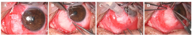
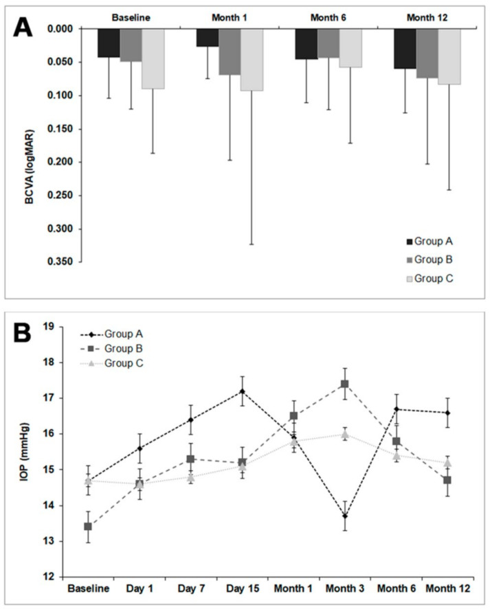
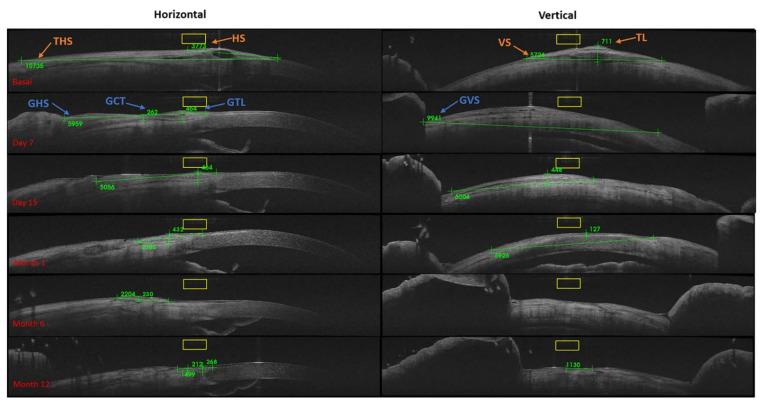
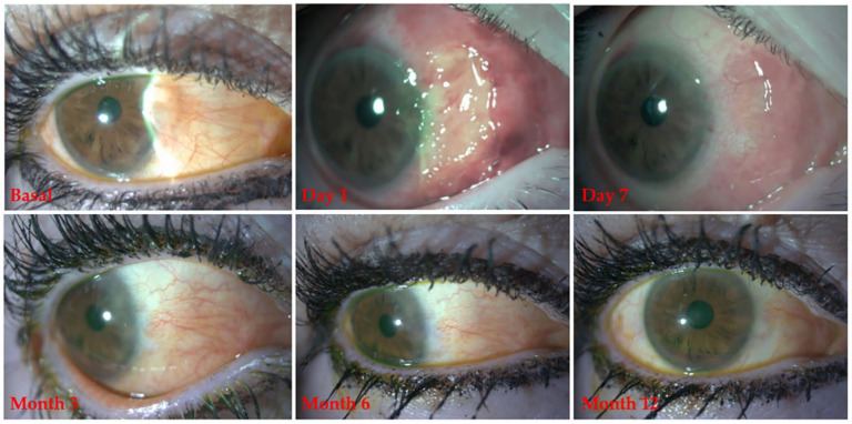
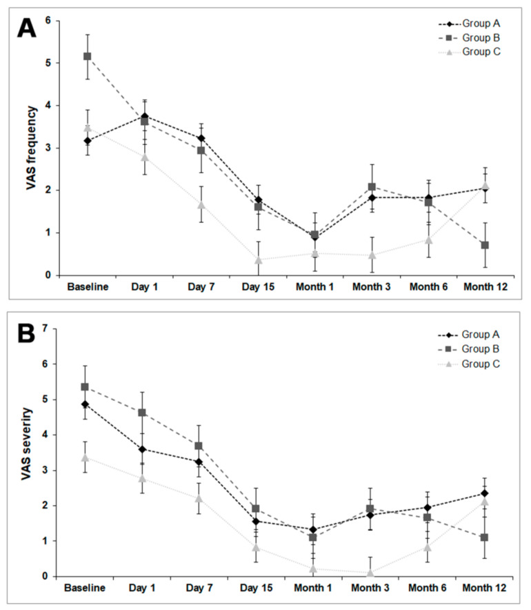
This prospective and comparative study aimed to compare the use of a conjunctival autograft (CAG), plasma rich in growth factors fibrin membrane (mPRGF) or amniotic membrane transplantation (AMT) in primary pterygium surgery. Patients were assigned for surgery with CAG (group A), mPRGF (group B), or AMT (group C). Pterygium recurrence, Best Corrected Visual Acuity (BCVA), graft size (measured with anterior segment optical coherence tomography (AS-OCT)), and ocular surface symptoms (visual analogue scale (VAS) and ocular surface disease index (OSDI)) were evaluated. Thirteen eyes in group A, 26 in group B, and 10 in group C were evaluated. No changes in BCVA (p > 0.05) were found. Recurrence cases for groups A, B, and C were none, two, and two, respectively, and three cases of pyogenic granulomas in group A. The horizontal/vertical graft size was lower in group B vs group A (p < 0.05) from months 1 to 12. The improvement in VAS frequency for groups A, B, and C was: 35.5%, 86.2%, and 39.1%, respectively. The OSDI scale reduction for groups A, B, and C was: 12.7%, 39.0%, and 84.1%. The use of the three surgical techniques as a graft for primary pterygium surgery was safe and effective, showing similar results. The mPRGF graft represents an autologous novel approach for pterygium surgery.
Keywords: PRGF; PRP; amniotic membrane transplantation; conjunctival autograft; plasma rich in growth factors; pterygium surgery.
Figures